Pupillary Light Reflex and Healthy Aging: What the Eyes Reveal
The pupil's response to light changes with age, medication, and eye health. Learn what pupillometry measures and why it is not a diagnosis.
Table of Contents
DISCLAIMER
This article is for informational purposes only and does not constitute medical advice. The statements in this article have not been evaluated by the FDA. The information presented is based on published research and should not be used as a substitute for professional medical guidance. Consult your physician before starting any supplement or health protocol.
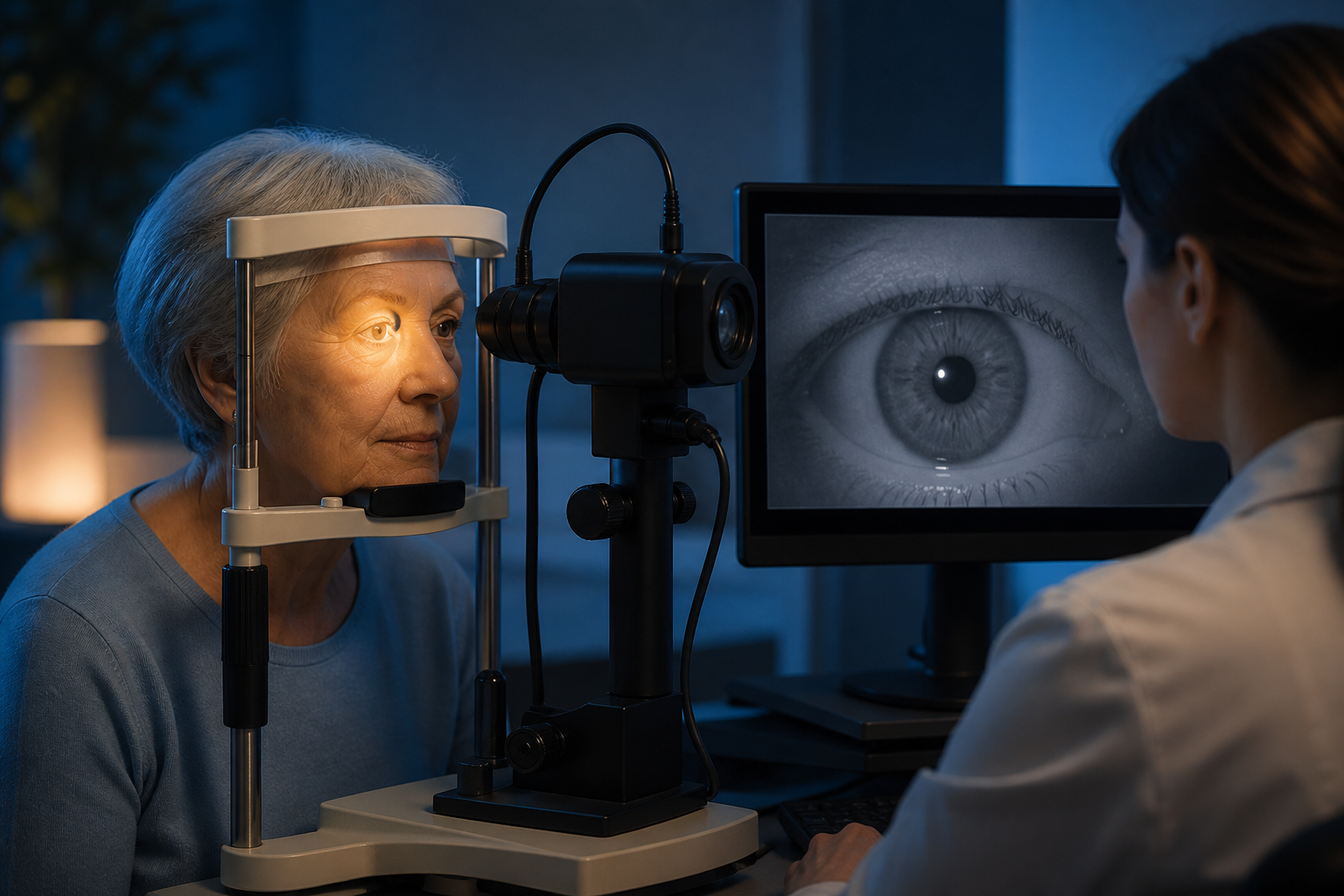
The pupil is a moving aperture, not a fixed black circle. When light reaches the retina, the pupil normally constricts; when the scene darkens, it dilates. The pupillary light reflex links the eye to brainstem and autonomic pathways, which makes it scientifically interesting—but not a simple meter of biological age.
Healthy aging can change resting pupil size and the speed or extent of its response. Those changes overlap with normal variation, eye disease, medication effects, and differences in measurement.
How the Light Reflex Works
Light-sensitive retinal cells send signals through the optic nerve toward midbrain centers. Each eye contributes to a shared circuit, so shining light in one eye normally constricts both the illuminated pupil and the opposite pupil.
Parasympathetic pathways drive constriction through the iris sphincter muscle. Sympathetic pathways help dilation through a different iris muscle. The final movement therefore reflects the stimulus, retinal input, neural transmission, iris mechanics, and the balance of autonomic activity.
Clinicians observe pupil size, symmetry, shape, and reaction as part of a broader examination. Laboratory pupillometry adds timing: latency before constriction, maximum constriction, velocity, redilation, and response to repeated flashes.
What Commonly Changes With Age
Older adults often have smaller pupils in darkness, a phenomenon sometimes called senile miosis. The maximum excursion can be reduced, while responses may be slower. Age-related iris stiffness, changes in autonomic signaling, and differences in retinal illumination may all contribute.
The lens also becomes denser with age, altering how much and which wavelengths of light reach the retina. Cataract, prior eye surgery, retinal disease, and glaucoma can affect measurements. A result that differs from a younger reference group is therefore not automatically abnormal.
Medications complicate interpretation. Some eye drops directly constrict or dilate the pupil; other prescription and nonprescription drugs can influence autonomic tone. Caffeine, nicotine, alertness, emotional arousal, and recent light exposure may shift a recording.
How Researchers Measure the Reflex
Dynamic pupillometers use infrared cameras so the pupil can be recorded without adding visible light. A standardized flash is delivered after a defined period of adaptation. Software tracks the pupil boundary frame by frame.
Small setup differences matter. Camera angle, eyelid position, room brightness, stimulus intensity, distance, and calibration affect the curve. Dark irises and reflections can challenge segmentation algorithms, while glasses or contact lenses may introduce artifacts.
Researchers are studying pupil responses in neurological, metabolic, sleep, and eye conditions. Promising group differences do not establish a consumer screening test. Many studies are small, cross-sectional, or use devices whose values cannot be compared directly.
What a Pupil Reading Can—and Cannot—Say
The reflex can help localize a problem when interpreted by a trained clinician alongside vision, eye movements, symptoms, and neurological findings. It cannot by itself identify a disease, measure longevity, or separate normal aging from every possible cause of a slow response.
Phone cameras have encouraged experimental home tools, but automatic exposure and screen brightness are not laboratory stimuli. Frame rate may be too low for accurate latency, and an app may not account for medication, cataract, or adaptation. The output should not be treated as an autonomic-health score.
A long-standing small difference between pupils can be benign. A sudden new difference is different—particularly with drooping eyelid, severe headache, eye pain, double vision, confusion, or weakness. Those combinations need prompt assessment rather than self-testing.
Supporting Vision Across Aging
Regular comprehensive eye examinations remain more useful than optimizing a pupil metric. Lighting at home can be improved because a smaller dark-adapted pupil reduces the light available in dim settings. Higher contrast on stairs and reading surfaces may help everyday function.
Sleep, activity, and cardiovascular risk management support general neurological and eye health, but no lifestyle routine should be claimed to normalize the light reflex. The goal is functional vision and timely care, not a particular millimeter value.
The Bottom Line
The pupillary light reflex is an elegant window into retinal, brainstem, iris, and autonomic function. Aging often changes the response, yet careful standardization and clinical context are essential; a camera reading alone cannot diagnose health or define how well someone is aging.
Frequently Asked Questions
Do pupils normally become smaller with age?
Can a camera app test autonomic health?
When does a pupil difference need urgent care?
Sources
Stay Updated on Longevity Science
Weekly research digests. No spam, unsubscribe anytime.
Related Articles
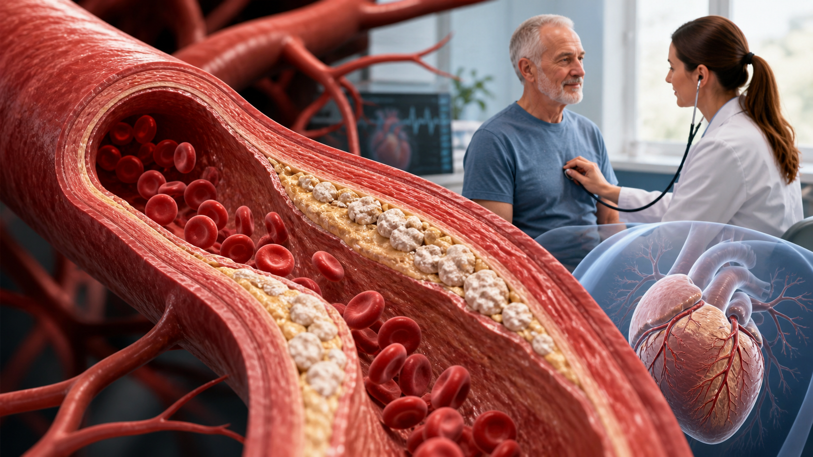
Arterial Calcification and Vascular Aging: Reading the Risk Signals
Arterial calcification can reveal accumulated vascular risk. Learn what calcium scores mean, why calcification develops, and where testing has limits.
10 min read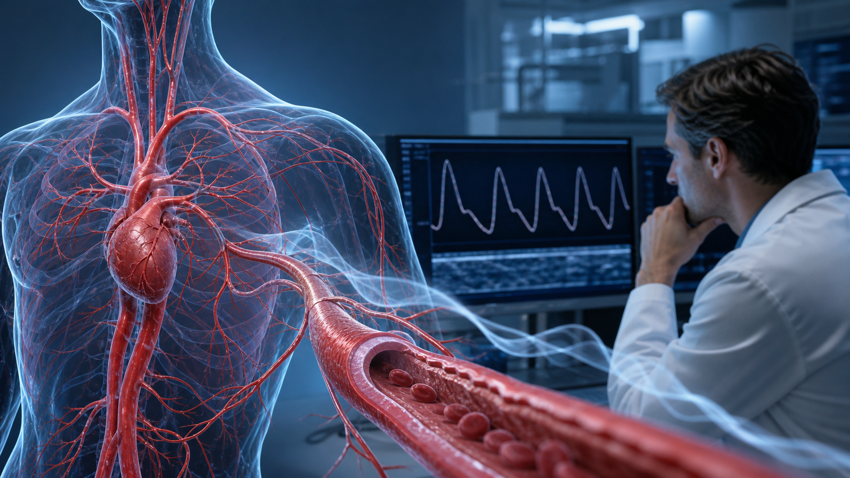
Arterial Stiffness and Vascular Age: A Longevity Evidence Guide
Understand arterial stiffness vascular age longevity links, key studies, measurement limits, and evidence-based lifestyle implications for healthy aging.
11 min read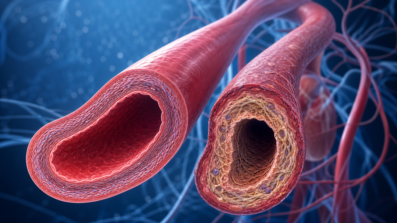
Arterial Stiffness and Vascular Aging: What the Evidence Shows
Explore arterial stiffness vascular aging, how vessels change over time, what pulse wave velocity studies show, and practical ways evidence may inform prevention.
10 min read