Mitophagy and Aging: How Cells Retire Damaged Mitochondria
Mitophagy and aging are linked through cellular quality control that removes damaged mitochondria. Learn what current evidence shows and what remains uncertain.
Table of Contents
DISCLAIMER
This article is for informational purposes only and does not constitute medical advice. The statements in this article have not been evaluated by the FDA. The information presented is based on published research and should not be used as a substitute for professional medical guidance. Consult your physician before starting any supplement or health protocol.
Mitophagy cellular quality control aging research asks a practical biological question: what happens when the tiny energy-producing structures inside cells wear out? Mitochondria continually experience stress as they convert nutrients into usable energy. A healthy cell does not simply keep every mitochondrion forever. It repairs some, merges others, divides networks into smaller units, and removes components that are no longer safe or useful.
That selective removal process is called mitophagy. It may help prevent damaged mitochondria from accumulating, producing excessive reactive molecules, or sending abnormal inflammatory signals. Mitophagy is therefore often discussed as a possible bridge between mitochondrial health and healthy aging. The evidence is promising, but it does not support treating mitophagy as a single switch that determines lifespan.
What Mitophagy Actually Does
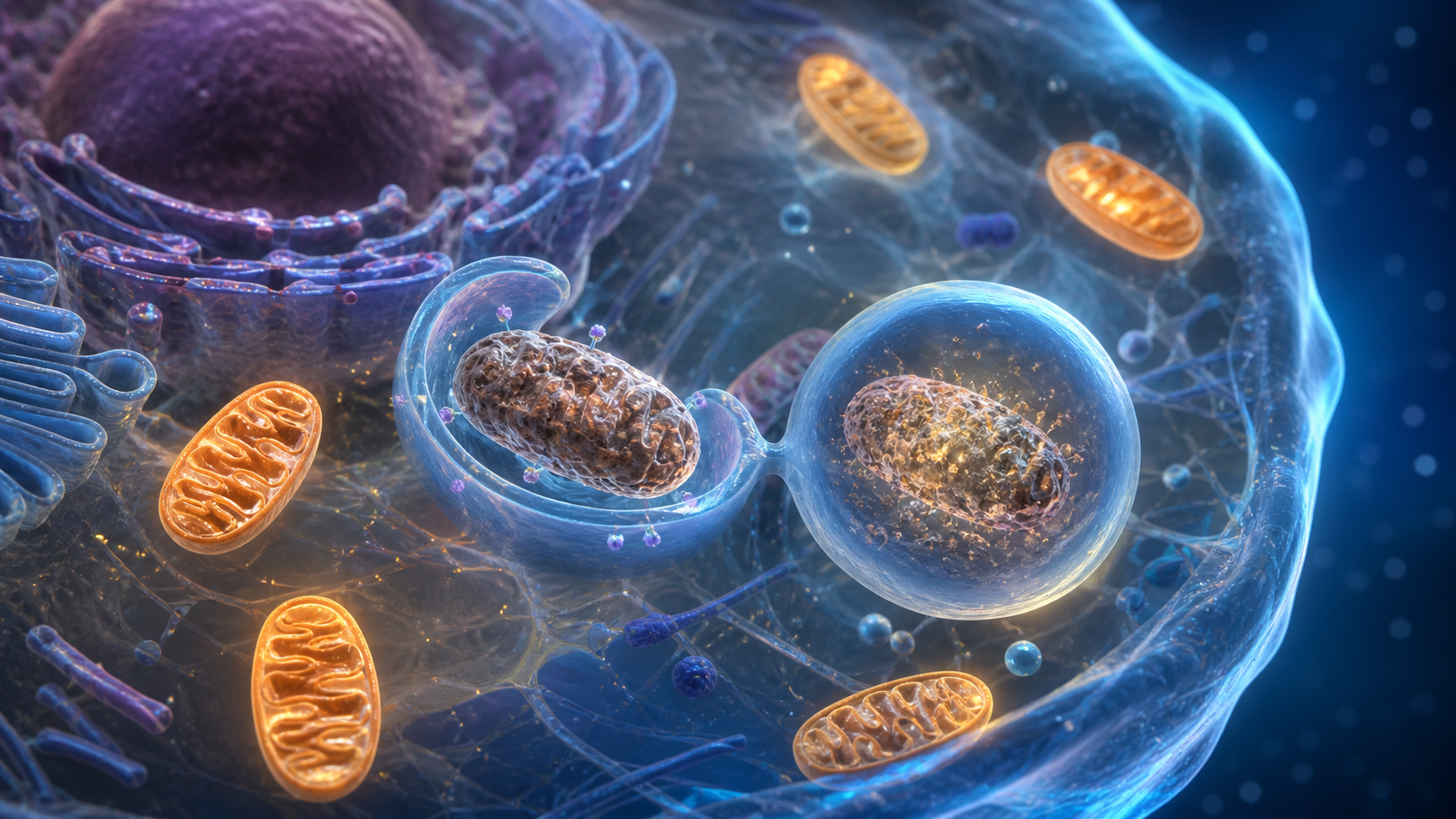
Mitophagy is a specialized form of autophagy. Autophagy broadly describes how cells package worn proteins, membranes, and organelles for delivery to lysosomes, where the material can be broken down and reused. Mitophagy applies this quality-control logic specifically to mitochondria.
Mitochondria operate as dynamic networks. They undergo fusion, which can mix contents and temporarily compensate for local damage, and fission, which can separate a troubled section from the healthier network. A fragment with persistent loss of membrane potential or other damage may then be tagged for removal.
One of the best-studied routes involves PINK1 and Parkin. Under ordinary conditions, the PINK1 protein is imported into healthy mitochondria and rapidly degraded. When a mitochondrion loses membrane potential, PINK1 can accumulate on its outer membrane. It then helps recruit and activate Parkin, an enzyme that adds molecular tags to mitochondrial proteins. Adapter proteins connect those tags to the membrane that forms an autophagosome. The compartment eventually fuses with a lysosome, completing degradation.
Cells also use Parkin-independent pathways. Receptors such as BNIP3, NIX, and FUNDC1 may link mitochondria more directly to autophagy machinery, particularly during low oxygen, red blood cell maturation, or tissue-specific stress. This redundancy matters: mitophagy is not one pathway, and different tissues may rely on different routes.
Why Mitochondrial Quality Control May Change With Age
Several parts of mitochondrial maintenance may become less coordinated with age. Lysosomal acidity and enzyme activity can change. Autophagy initiation may weaken. Mitochondrial fission and fusion may become imbalanced. Damaged proteins and mitochondrial DNA variants may accumulate. At the same time, chronic inflammation, inactivity, metabolic disease, and medication exposure can alter the same systems.
The result is not necessarily a total failure of mitophagy. Aging is heterogeneous, and cells may compensate for years. The more useful model is declining reserve: a tissue may manage ordinary damage but struggle when infection, surgery, prolonged inactivity, or another metabolic challenge sharply raises demand.
Muscle and neurons make the issue especially visible. Both depend heavily on energy and must maintain large, spatially complex mitochondrial networks. Neurons cannot easily replace themselves, while muscle function depends on coordinated energy delivery across long fibers. A backlog of dysfunctional mitochondria may therefore influence movement, cognition, and resilience even when it does not cause a specific disease by itself.
What Key Research Findings Suggest
Youle and Narendra’s 2011 review helped organize the molecular evidence around PINK1, Parkin, mitochondrial damage sensing, and selective removal. Much of the foundational work involved genetic models connected to Parkinson’s disease. These models demonstrated that defects in mitochondrial quality control can matter biologically, but they do not show that activating the pathway indiscriminately would benefit every person.
Palikaras and colleagues reported in 2015 that mitophagy and mitochondrial biogenesis are coordinated during aging in C. elegans. Their work linked mitochondrial turnover with stress signaling and lifespan in a simple organism. The study supports the principle that removal and replacement need to be balanced. Clearing mitochondria without rebuilding capacity would not create a healthy energy system.
Ryu and colleagues reported in 2016 that urolithin A induced mitophagy in laboratory models, extended lifespan in worms, and improved some measures of muscle function in rodents. The result attracted interest because gut microbes can produce urolithins from compounds found in foods such as pomegranates and walnuts. Translation remains challenging: microbial conversion differs between people, animal outcomes do not guarantee human outcomes, and supplement trials must evaluate meaningful functional endpoints rather than pathway markers alone.
Human studies increasingly measure mitochondrial turnover signals in blood or muscle, but direct measurement remains difficult. A rise in one protein does not prove that the entire removal process reached completion. Researchers distinguish autophagosome formation from autophagic flux—the successful movement of material through degradation. A traffic jam can create many delivery vehicles without completing more deliveries.
How Exercise, Sleep, and Metabolic Health Fit In
Regular exercise is one of the strongest real-world stimuli for mitochondrial remodeling. Endurance activity can challenge energy production, while resistance exercise creates mechanical and metabolic signals in muscle. Recovery then supports repair, biogenesis, and turnover. Research suggests mitophagy-related signaling participates in this adaptation, although the exact response depends on exercise dose and sampling time.
This does not mean harder is always better. Excess training without recovery can add damage faster than a person can adapt. Older adults, people with cardiovascular or neurological conditions, and anyone returning after long inactivity may benefit from gradual progression and professional guidance.
Sleep and circadian timing may also matter because autophagy, metabolism, and hormone signals follow daily rhythms. Evidence supports consistent sleep as part of general metabolic health, but no consumer sleep schedule has been proven to optimize human mitophagy directly.
Dietary energy balance affects nutrient-sensing pathways such as AMPK and mTOR that interact with autophagy. Fasting experiments can activate cellular recycling signals in animals, yet the timing and magnitude in humans are variable. Aggressive fasting can be risky for people who are frail, underweight, pregnant, taking glucose-lowering medication, or recovering from illness. A pathway observation should not be turned into universal medical advice.
Limitations and Future Research
The largest limitation is translation. Worm lifespan, mouse treadmill performance, and cultured-cell markers answer important questions, but not the final human question: does a safe intervention preserve independence or reduce disease over years? Human tissues are difficult to sample repeatedly, and mitophagy changes quickly after meals, exercise, sleep, and stress.
Researchers also need tissue-specific answers. Increasing mitophagy in muscle may not have the same effect in immune cells, liver, heart, or brain. Too little removal can leave damaged components in place, but too much removal without replacement may reduce mitochondrial capacity. The goal is efficient turnover, not maximal destruction.
Future trials may combine imaging, muscle biopsies, stable-isotope tracing, circulating markers, and functional outcomes. They will also need to separate the effects of a candidate compound from the fundamentals that influence mitochondrial health: physical activity, smoking exposure, sleep, metabolic disease, and nutrition.
The Bottom Line
Mitophagy is a credible part of the biology of aging because it helps cells identify and recycle damaged mitochondria. Laboratory evidence suggests that preserving this quality-control system may support tissue function, but no single mitophagy booster has been shown to reverse human aging.
For now, the most defensible interpretation is that mitochondrial health depends on balanced maintenance: damage sensing, selective removal, replacement, and appropriate energy demand. People considering supplements or major fasting or exercise changes should consult a qualified healthcare provider, especially when medical conditions or medications are involved.
Frequently Asked Questions
What is mitophagy?
Does mitophagy slow human aging?
Can exercise influence mitophagy?
Sources
Stay Updated on Longevity Science
Weekly research digests. No spam, unsubscribe anytime.
Related Articles
Arterial Calcification and Vascular Aging: Reading the Risk Signals
Arterial calcification can reveal accumulated vascular risk. Learn what calcium scores mean, why calcification develops, and where testing has limits.
10 min read
Arterial Stiffness and Vascular Age: A Longevity Evidence Guide
Understand arterial stiffness vascular age longevity links, key studies, measurement limits, and evidence-based lifestyle implications for healthy aging.
11 min read
Arterial Stiffness and Vascular Aging: What the Evidence Shows
Explore arterial stiffness vascular aging, how vessels change over time, what pulse wave velocity studies show, and practical ways evidence may inform prevention.
10 min read