Contrast Sensitivity and Healthy Aging: Vision Beyond the Eye Chart
Contrast sensitivity can decline even when standard visual acuity looks normal. Learn how aging affects real-world vision, testing, mobility, and safety.
Table of Contents
DISCLAIMER
This article is for informational purposes only and does not constitute medical advice. The statements in this article have not been evaluated by the FDA. The information presented is based on published research and should not be used as a substitute for professional medical guidance. Consult your physician before starting any supplement or health protocol.
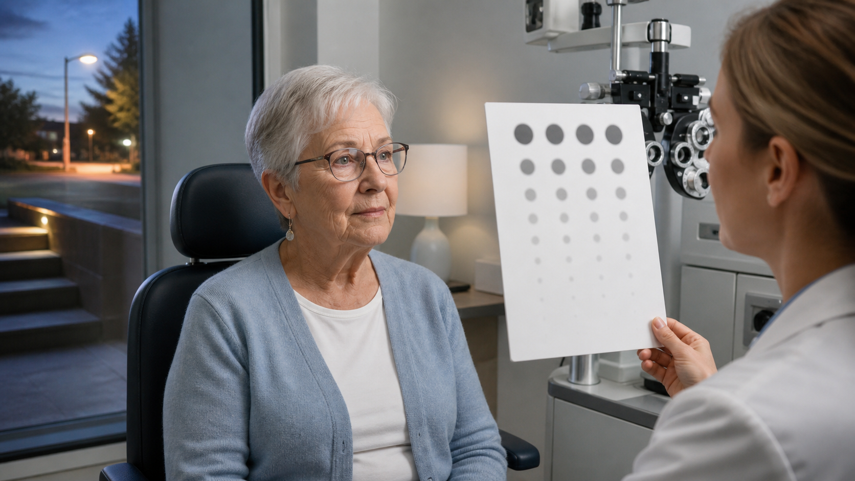
Contrast sensitivity in vision and healthy aging describes something a standard eye chart can miss: how well the visual system separates an object from a similar background. Black letters on white paper are a high-contrast task. A gray curb at dusk, a face in a dim restaurant, or a step in shadow is not.
This difference matters because everyday vision rarely arrives in perfect laboratory conditions. Aging can reduce the amount and quality of light reaching the retina, increase optical scatter, and change neural processing. A person may still read the smallest chart line while feeling less secure at night.
How Contrast Sensitivity Differs From Acuity
Visual acuity asks how small a high-contrast detail can be resolved. Contrast sensitivity asks how faint a pattern can be detected across different sizes. The resulting contrast-sensitivity function provides a broader picture of visual performance.
Large, blurry objects occupy low spatial frequencies; fine details occupy high spatial frequencies. Different eye diseases and optical problems can affect different parts of this range. A single acuity score compresses that complexity.
Contrast testing is sensitive to room lighting, glare, refractive correction, chart type, and test distance. This makes standardization important. It also means a consumer screen test cannot replace a complete eye examination.
Why aging changes the signal
The pupil often becomes smaller with age, allowing less light to reach the retina in dim conditions. The crystalline lens can yellow and scatter more light, even before a cataract is considered visually significant. Tear-film instability and uncorrected refractive error can reduce image quality.
The retina and visual pathways also change. Photoreceptor function, retinal circuitry, and cortical processing all contribute to detecting contrast. Healthy aging may therefore affect both the optical image and the brain’s interpretation of it.
What Research Connects to Daily Function
Lower contrast sensitivity has been associated with difficulty in mobility, face recognition, reading under poor conditions, and driving—especially at night or in glare. Observational research has also connected several measures of visual function with falls and fractures.
These associations require caution. Falls have many causes, and people with visual impairment may also differ in balance, medication use, health conditions, or activity exposure. Contrast sensitivity is one risk signal, not a complete prediction.
The practical value is that visual acuity alone may underestimate difficulty. If someone reports that steps disappear in shadows or headlights make the road unreadable, “normal” chart acuity should not end the conversation.
Eye disease and contrast loss
Cataract can scatter light and reduce contrast, particularly in glare. Glaucoma may affect spatial vision even before a person notices central blur. Macular disease can disrupt fine detail and contrast, while diabetic eye disease can affect multiple retinal layers.
Because these conditions have different management pathways, a contrast result should prompt appropriate examination rather than self-diagnosis. Sudden vision loss, flashes, a curtain-like shadow, new double vision, or eye pain needs urgent professional attention.
Healthy-Aging Implications
Regular comprehensive eye care becomes more important with age because several eye diseases can progress quietly. Updating a prescription, managing dry eye, treating cataract when appropriate, and monitoring retinal or optic-nerve disease may improve or protect real-world function.
Environment can reduce unnecessary visual demand. Even lighting, clear edge contrast on stairs, glare control, and uncluttered walkways may help a person use the vision available. These are not substitutes for diagnosis; they are design choices that make hazards easier to see.
Mobility training also matters. People sometimes reduce activity when vision feels uncertain, which may lower strength and confidence. A coordinated plan can address both the visual limitation and the physical capacities needed for safe movement.
Limits of a “Vision Age”
Contrast sensitivity is tempting as a biological-age marker because it changes across adulthood and connects to function. But the result can be altered by treatable eye conditions, testing conditions, fatigue, and equipment. It does not isolate one aging mechanism.
Longitudinal change is more informative than one score, provided the method is consistent. Even then, interpretation belongs beside acuity, visual fields, retinal and optic-nerve findings, symptoms, and daily function.
The Bottom Line
Contrast sensitivity reveals a dimension of healthy vision that the familiar black-and-white chart can overlook. It may help explain problems in dim light, glare, mobility, and driving, but it is neither a standalone diagnosis nor a whole-body aging clock. The useful goal is better real-world function, not a younger-looking number.
Frequently Asked Questions
What is contrast sensitivity?
Can contrast sensitivity decline with normal 20/20 vision?
Does poor contrast sensitivity mean dementia?
Sources
Stay Updated on Longevity Science
Weekly research digests. No spam, unsubscribe anytime.
Related Articles
Arterial Calcification and Vascular Aging: Reading the Risk Signals
Arterial calcification can reveal accumulated vascular risk. Learn what calcium scores mean, why calcification develops, and where testing has limits.
10 min read
Arterial Stiffness and Vascular Age: A Longevity Evidence Guide
Understand arterial stiffness vascular age longevity links, key studies, measurement limits, and evidence-based lifestyle implications for healthy aging.
11 min read
Arterial Stiffness and Vascular Aging: What the Evidence Shows
Explore arterial stiffness vascular aging, how vessels change over time, what pulse wave velocity studies show, and practical ways evidence may inform prevention.
10 min read