Blood-Brain Barrier Aging: What Changes and Why It Matters
Blood-brain barrier aging research links vascular changes with brain health, but causality is complex. Learn what human studies show and what remains unknown.
Table of Contents
DISCLAIMER
This article is for informational purposes only and does not constitute medical advice. The statements in this article have not been evaluated by the FDA. The information presented is based on published research and should not be used as a substitute for professional medical guidance. Consult your physician before starting any supplement or health protocol.
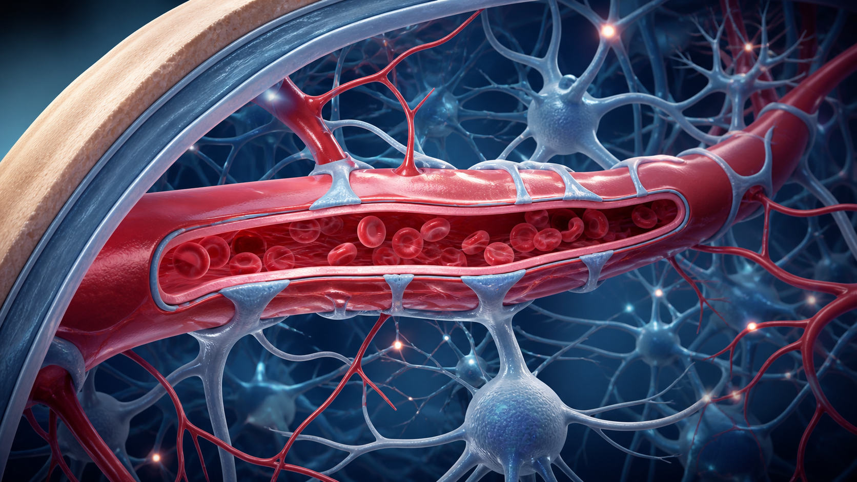
Blood brain barrier aging research examines the interface between circulation and the brain. The blood-brain barrier is often imagined as a wall, but it is better understood as a controlled border made from capillary endothelial cells, tight junctions, pericytes, astrocyte end-feet, basement membrane, and transport systems.
What the Barrier Protects
Brain cells require oxygen, glucose, amino acids, hormones, and carefully regulated ions. At the same time, uncontrolled entry of inflammatory molecules, toxins, or immune cells could disturb neural signaling. The barrier manages this tradeoff through selective transport, enzymes, efflux pumps, and physical junctions between endothelial cells.
It also belongs to the neurovascular unit. Blood flow, neurons, glial cells, and vessel walls communicate. A change in one component can affect the others, which is why barrier dysfunction is difficult to separate from wider vascular aging.
How Aging May Alter the Neurovascular Unit
With age, endothelial signaling may become less responsive, pericyte coverage may change, basement membranes may thicken, and chronic low-grade inflammation may influence tight junctions. Hypertension, diabetes, smoking, kidney disease, sleep disruption, and inactivity can add vascular stress.
Transport may change even without dramatic leakage. The brain may become less efficient at moving certain nutrients inward or clearing metabolites outward. These processes vary by brain region and disease state. The hippocampus, involved in memory, has received particular attention.
What Human Imaging Studies Found
Montagne and colleagues used dynamic contrast-enhanced MRI and cerebrospinal-fluid markers in 2015. They reported age-related barrier breakdown in the hippocampus, particularly in regions involved in learning and memory, and linked it with pericyte injury markers. The work provided human evidence that vascular change can appear during aging, but it did not show that leakage alone causes cognitive decline.
Nation and colleagues reported in 2019 that barrier breakdown in the hippocampus was associated with early cognitive dysfunction independently of conventional amyloid and tau measures in their cohort. This supported a vascular pathway that may run alongside better-known Alzheimer’s mechanisms. Cohort design, imaging thresholds, and participant selection still limit broad conclusions.
Reviews by Sweeney and colleagues describe a two-way system: vascular dysfunction can influence neurons, and neurological disease can further impair the barrier. This feedback makes timing crucial. A marker observed late in disease may be a driver, a consequence, or both.
What Supports Vascular Brain Health
No consumer routine has been proven to seal the blood-brain barrier. The strongest practical evidence instead supports managing established vascular risks. Blood-pressure care, diabetes management, avoiding smoking, regular physical activity, and treatment of sleep apnea can protect circulation broadly and may indirectly support the neurovascular unit.
Exercise can improve endothelial function and cerebral blood-flow regulation, but trials rarely measure barrier permeability as the main clinical endpoint. A healthy dietary pattern may support cardiometabolic health without acting as a direct barrier treatment.
Claims around fasting, ketones, flavonoids, or supplements often begin with cell or animal studies. Such findings may guide research but should not be presented as evidence of barrier repair in humans. Products may also interact with medication or create risk when they replace standard care.
Limitations, Future Research, and the Bottom Line
Barrier measurement is technically demanding. MRI contrast methods provide indirect estimates and may differ across scanners and analysis pipelines. Blood markers can originate outside the brain. Cerebrospinal-fluid sampling is invasive. Researchers need harmonized methods and longitudinal studies that connect barrier change with meaningful outcomes.
Future work may identify distinct vascular-aging profiles and test whether treating blood pressure, metabolic disease, inflammation, or pericyte injury changes the trajectory. It will also need to account for sex, genetics, ethnicity, medication, and coexisting disease.
The blood-brain barrier is a dynamic component of brain health, and evidence suggests that parts of this system can change with age. The science does not justify fear of a universally leaking brain or confidence in a quick repair. People concerned about memory change or vascular risk should consult a healthcare professional for evaluation and established risk management.
Frequently Asked Questions
What does the blood-brain barrier do?
Does the blood-brain barrier always weaken with age?
Can a supplement repair the blood-brain barrier?
Sources
Stay Updated on Longevity Science
Weekly research digests. No spam, unsubscribe anytime.
Related Articles
Arterial Calcification and Vascular Aging: Reading the Risk Signals
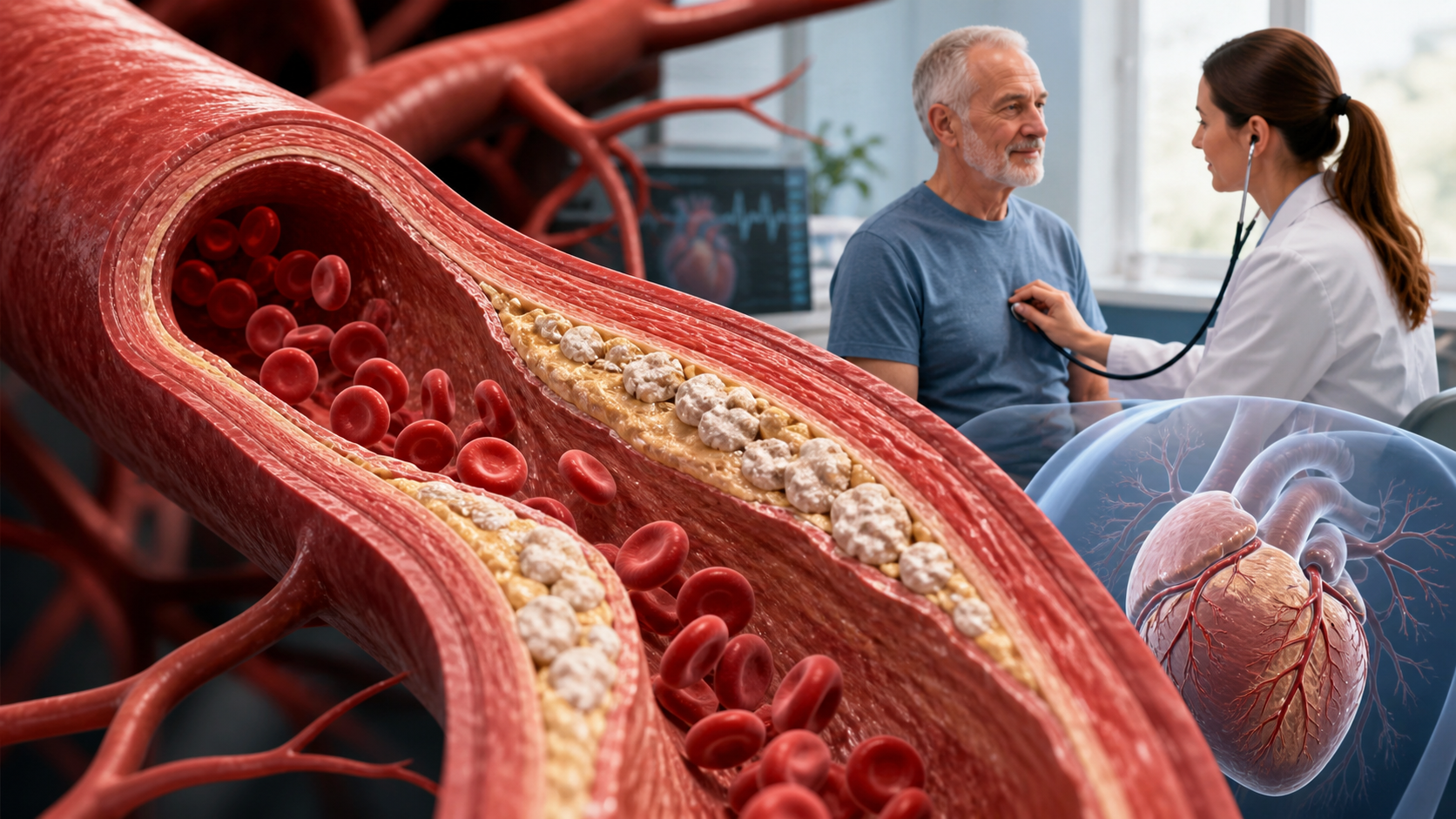
Arterial calcification can reveal accumulated vascular risk. Learn what calcium scores mean, why calcification develops, and where testing has limits.
10 min read
Arterial Stiffness and Vascular Age: A Longevity Evidence Guide
Understand arterial stiffness vascular age longevity links, key studies, measurement limits, and evidence-based lifestyle implications for healthy aging.
11 min read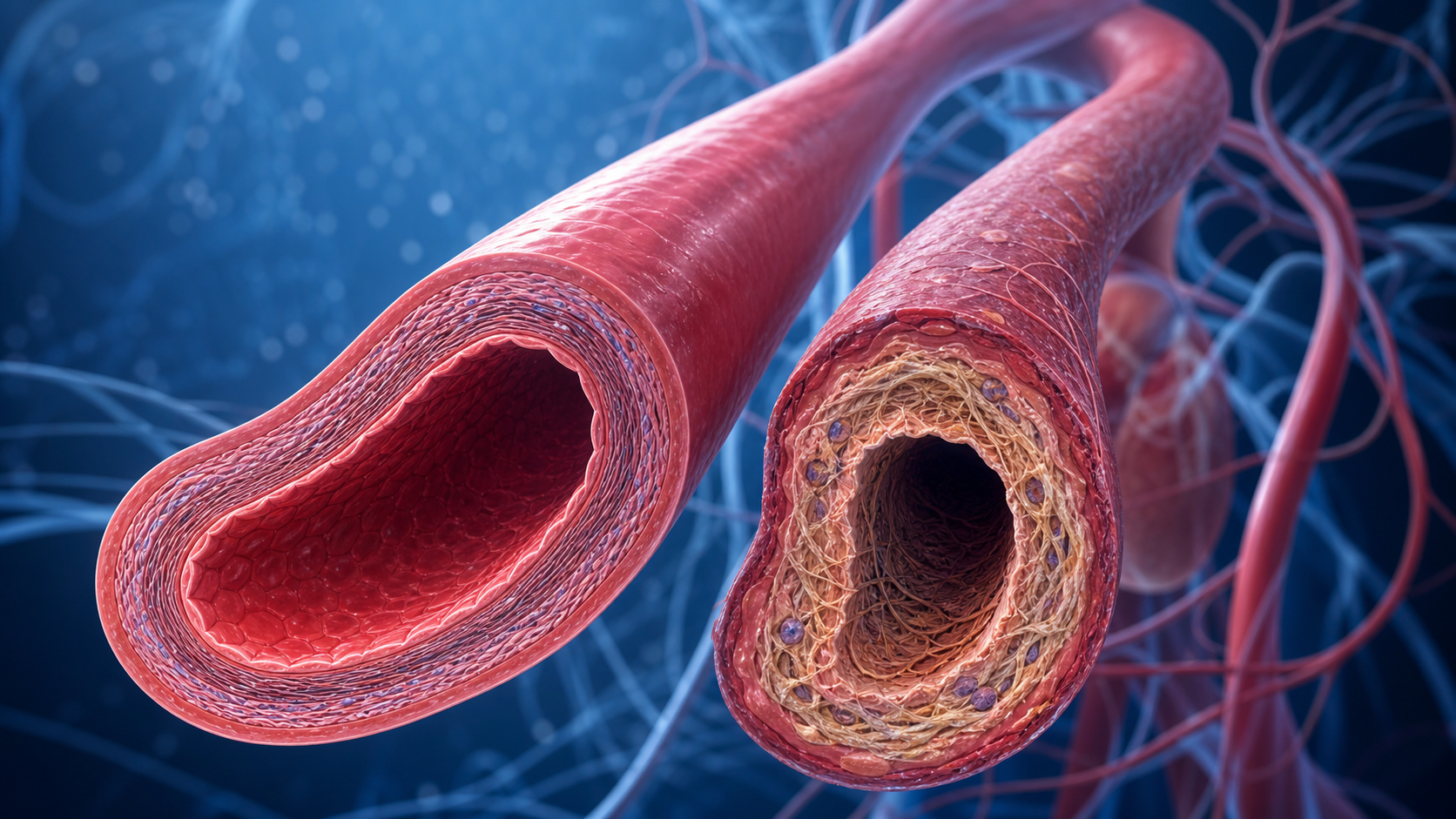
Arterial Stiffness and Vascular Aging: What the Evidence Shows
Explore arterial stiffness vascular aging, how vessels change over time, what pulse wave velocity studies show, and practical ways evidence may inform prevention.
10 min read