Autophagy Flux and Healthy Aging: What Biomarkers Can Actually Show
Autophagy recycles damaged cell material, but measuring it in people is difficult. Learn what autophagy flux means and how to interpret current biomarkers.
Table of Contents
DISCLAIMER
This article is for informational purposes only and does not constitute medical advice. The statements in this article have not been evaluated by the FDA. The information presented is based on published research and should not be used as a substitute for professional medical guidance. Consult your physician before starting any supplement or health protocol.
Autophagy is often described as the cell’s recycling system. That shorthand is useful, but it can hide the most important measurement problem: seeing more recycling bins does not prove that more waste was successfully processed.
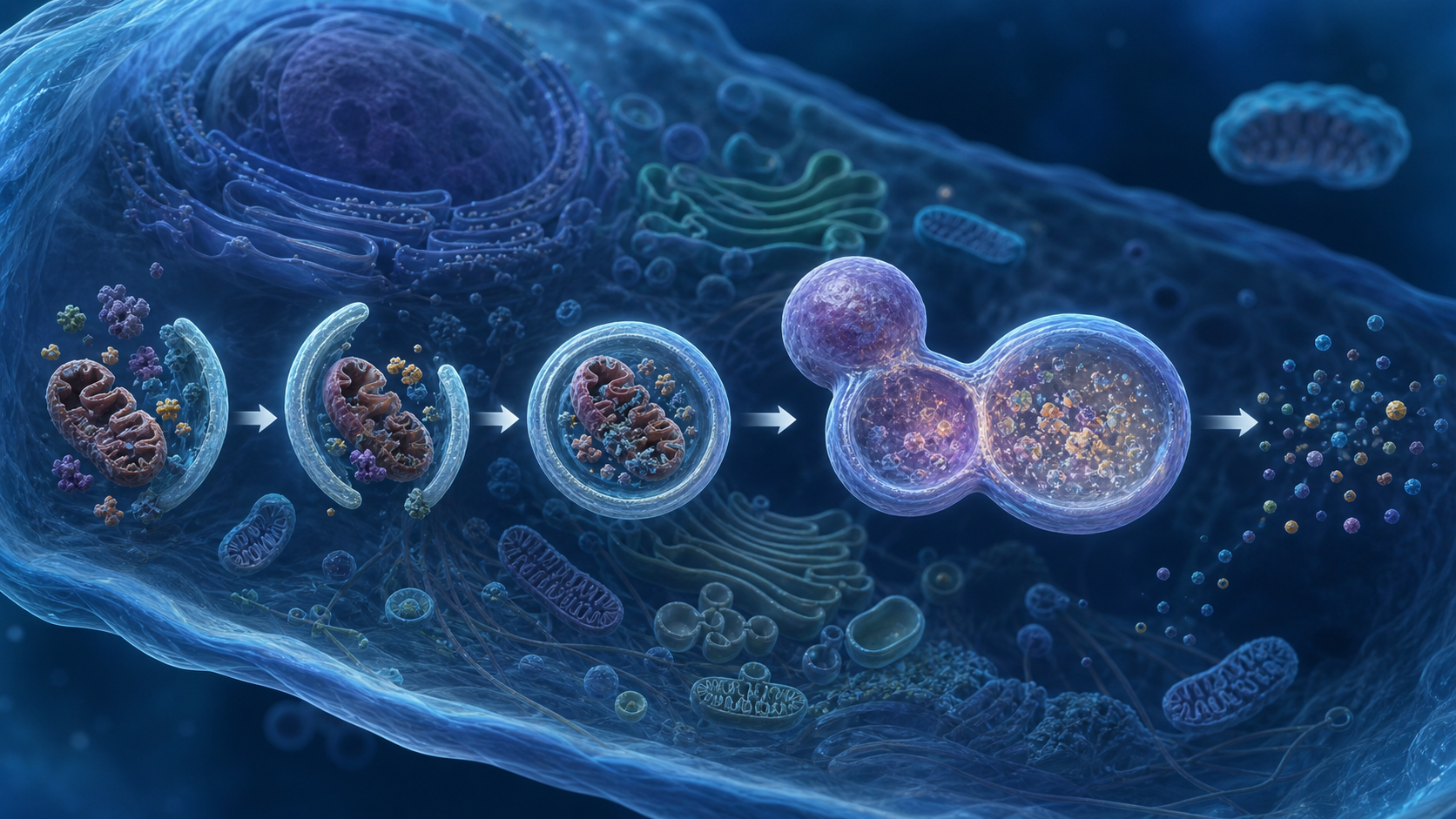
Researchers therefore focus on autophagy flux—the movement of material through the entire pathway. Flux includes formation of an autophagosome, delivery to a lysosome, breakdown of the cargo, and reuse or disposal of the resulting components.
What Autophagy Flux Means
Cells surround selected proteins, lipids, and organelle fragments with a double-membrane structure called an autophagosome. It then fuses with a lysosome, whose enzymes degrade the cargo. The pathway helps cells respond to nutrient stress and remove damaged material.
A snapshot can be misleading. Many autophagosomes may mean that the pathway is active. They may also mean that degradation is blocked and material is piling up. Flux asks whether cargo enters and exits the system over time.
This distinction is especially relevant to aging. Older tissues may show impaired lysosomal acidity, slower cargo recognition, altered nutrient sensing, or reduced capacity to finish the process. A useful assay must distinguish increased initiation from delayed clearance.
How Researchers Measure the Pathway
Laboratories commonly study proteins such as LC3 and p62/SQSTM1, image autophagic structures, or use tagged reporter systems. The strongest experiments compare measurements before and after lysosomal degradation is deliberately blocked. The difference helps estimate how much material was moving through the pathway.
These methods work best in cells, model organisms, or carefully collected tissue. Human studies face practical limits. A muscle biopsy samples one tissue at one moment. Blood cells are easier to collect but may not represent the brain, liver, muscle, or heart.
Gene-expression panels are even more indirect. More messenger RNA for an autophagy-related gene does not guarantee that the protein is active or that lysosomes complete degradation. Metabolites and extracellular vesicles may eventually add useful signals, but no single marker has become a validated consumer measure of whole-body flux.
Why Autophagy Is Linked to Healthy Aging
Autophagy tends to become less efficient in several aging models, while genetic or pharmacological changes that preserve parts of the pathway can extend lifespan in organisms such as yeast, worms, flies, and mice. The pathway intersects with proteostasis, mitochondrial quality control, immunity, and nutrient sensing.
Human evidence is more cautious. Exercise and energy restriction can influence upstream regulators such as AMPK and mTOR, but the response varies by tissue, training state, meal timing, and sampling method. Observational associations do not establish that increasing one marker will extend life.
Autophagy also has context-dependent effects. Cancer cells can use recycling to survive stress, while excessive or dysregulated pathway activity can accompany tissue injury. The goal is not permanent maximal activation. It is responsive cellular housekeeping that turns on and off appropriately.
How to Interpret Autophagy Claims
Be skeptical of products that promise a precise autophagy score from a routine blood draw. Ask which tissue was measured, whether the assay captured flux rather than a static marker, and whether the result predicts a meaningful clinical outcome.
Lifestyle evidence is strongest when it supports broader health outcomes. Regular physical activity, adequate sleep, and a nutritionally sound eating pattern can improve metabolic and cardiovascular health even when an individual’s autophagy response is not measured. Prolonged fasting or unapproved drugs can carry real risks and should not be treated as do-it-yourself pathway tests.
Autophagy is biologically important, but the honest conclusion is narrower than the marketing: scientists can measure flux well in controlled systems, while practical human biomarkers remain an active research problem.
Frequently Asked Questions
Can a blood test measure whole-body autophagy?
Does more autophagy always mean healthier aging?
Can fasting prove that my autophagy increased?
Sources
Stay Updated on Longevity Science
Weekly research digests. No spam, unsubscribe anytime.
Related Articles
Arterial Calcification and Vascular Aging: Reading the Risk Signals
Arterial calcification can reveal accumulated vascular risk. Learn what calcium scores mean, why calcification develops, and where testing has limits.
10 min read
Arterial Stiffness and Vascular Age: A Longevity Evidence Guide
Understand arterial stiffness vascular age longevity links, key studies, measurement limits, and evidence-based lifestyle implications for healthy aging.
11 min read
Arterial Stiffness and Vascular Aging: What the Evidence Shows
Explore arterial stiffness vascular aging, how vessels change over time, what pulse wave velocity studies show, and practical ways evidence may inform prevention.
10 min read