Brain Pericytes and Aging: Guardians of the Blood-Brain Barrier
Pericytes support brain capillaries and the blood-brain barrier. Explore how these cells may change with age, what studies suggest, and why the evidence needs context.
Table of Contents
DISCLAIMER
This article is for informational purposes only and does not constitute medical advice. The statements in this article have not been evaluated by the FDA. The information presented is based on published research and should not be used as a substitute for professional medical guidance. Consult your physician before starting any supplement or health protocol.
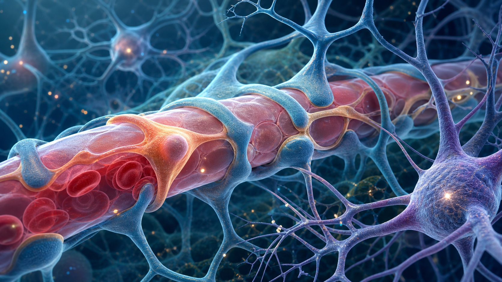
Every thought depends on a delivery system. The brain requires continuous oxygen and glucose, yet it must tightly control what crosses from blood into neural tissue. That balancing act belongs to the neurovascular unit: endothelial cells, pericytes, astrocytes, neurons, immune cells, and extracellular matrix working together around tiny vessels.
Pericytes are easy to overlook. These slender cells wrap portions of capillaries and small venules, sharing a basement membrane with endothelial cells. Research suggests that they help stabilize vessels, influence blood flow, support blood-brain barrier properties, and coordinate repair. Changes in pericyte coverage or signaling may therefore be one piece of brain aging, but the biology is still being mapped.
Where Pericytes Fit in the Blood-Brain Barrier
Brain endothelial cells form unusually tight junctions and use selective transport systems. Pericytes do not create those junctions directly, but experiments suggest that they help endothelial cells maintain the specialized gene-expression program required for barrier function.
Communication travels both ways. Endothelial cells release platelet-derived growth factor-B, which helps recruit and maintain pericytes through PDGFR-beta signaling. Pericytes release signals that influence endothelial survival, basement-membrane organization, and astrocyte interactions. Disturbing one cell type can change the entire unit.
Pericytes may also participate in capillary diameter control, although the size and timing of their contribution remain debated. Smooth-muscle cells clearly regulate larger arterioles. At the capillary level, contractile behavior, cell subtype, and experimental definitions make conclusions more difficult.
Another role is waste and immune regulation. Pericytes interact with pathways involved in clearance around vessels and can respond to inflammatory signals. They are not passive wrapping; they are positioned where metabolism, circulation, and immune surveillance meet.
What Changes With Age
Animal studies and human tissue research suggest that pericyte density, coverage, or function may decline in some brain regions with age. The degree varies by species, vascular segment, disease burden, and measurement method. A lower marker signal does not always mean that cells have disappeared; their molecular identity may have changed.
Blood-brain barrier permeability also appears to increase in certain older populations and brain regions. Imaging research has reported age-related leakage signals, including in the hippocampal area, while fluid studies have explored markers linked with pericyte injury. These findings support a vascular contribution to cognitive aging, but they do not establish a simple sequence in every person.
Barrier disruption could expose neural tissue to plasma proteins and inflammatory signals. In turn, inflammation and oxidative stress could impair pericytes and endothelial cells. Reduced perfusion may compromise energy delivery, while altered clearance may allow metabolites to accumulate. These loops make it difficult to identify a single starting point.
Common vascular risk factors complicate the picture. Hypertension, diabetes, smoking, kidney disease, and prior stroke can influence small vessels. Neurodegenerative disease can also affect the neurovascular unit. Studies must separate chronological age from the conditions that become more common with age.
How to Interpret the Research
There is no routine pericyte-health score. Dynamic contrast imaging, cerebrospinal-fluid markers, and soluble PDGFR-beta measurements are research tools, not stand-alone consumer diagnostics. Results can vary with technique and may reflect several tissues or processes.
The most defensible practical lesson is that brain health and vascular health are connected. Managing established cardiovascular risks with appropriate clinical guidance supports the circulation that the neurovascular unit depends on. Physical activity, sleep, hearing and vision care, social engagement, and metabolic health all belong to a broader brain-health strategy, but none can be claimed to target pericytes alone.
New neurological symptoms deserve prompt assessment. Sudden weakness, speech difficulty, severe headache, or confusion can indicate an emergency. Gradual memory changes also merit evaluation because medication effects, sleep disorders, mood, hearing loss, metabolic problems, and neurological disease may overlap.
Pericyte research offers a valuable correction to neuron-only stories of brain aging. The brain is an organ supplied by living vessels, and its smallest vascular cells help maintain the environment in which neurons work. The evidence suggests that pericytes matter; it does not yet justify a commercial test, a single-cause explanation for cognitive decline, or a promise that one intervention can seal the aging blood-brain barrier.
Frequently Asked Questions
What do brain pericytes do?
Does blood-brain barrier leakage mean dementia?
Can people directly test pericyte health?
Sources
Stay Updated on Longevity Science
Weekly research digests. No spam, unsubscribe anytime.
Related Articles

Ankle Power and Healthy Aging: The Push-Off That Keeps Walking Efficient
Ankle plantar-flexor power supports walking speed and balance recovery. Learn why push-off changes with age and how strength can be trained safely.
9 min read
Arterial Calcification and Vascular Aging: Reading the Risk Signals
Arterial calcification can reveal accumulated vascular risk. Learn what calcium scores mean, why calcification develops, and where testing has limits.
10 min read
Arterial Stiffness and Vascular Age: A Longevity Evidence Guide
Understand arterial stiffness vascular age longevity links, key studies, measurement limits, and evidence-based lifestyle implications for healthy aging.
11 min read