Retinal Microvasculature and Biological Aging
How retinal vessel measurements and fundus-image models may reflect vascular and biological aging, plus the evidence gaps that limit individual interpretation.
Table of Contents
DISCLAIMER
This article is for informational purposes only and does not constitute medical advice. The statements in this article have not been evaluated by the FDA. The information presented is based on published research and should not be used as a substitute for professional medical guidance. Consult your physician before starting any supplement or health protocol.
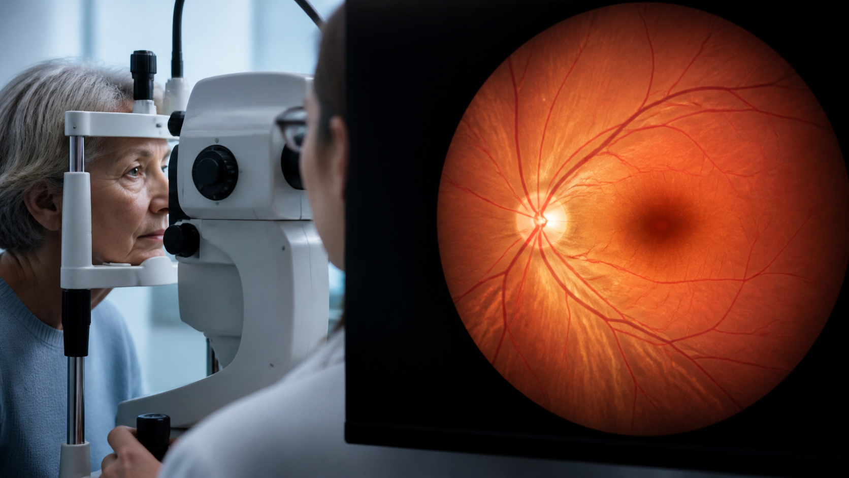
The retina offers something rare in human biology: a direct, noninvasive view of living blood vessels and neural tissue. A routine fundus photograph can reveal arteries, veins, branching patterns, hemorrhages, and features of the optic nerve without surgery. That accessibility has made retinal imaging a promising source of biomarkers for vascular and biological aging.
Machine-learning systems can often estimate chronological age from retinal photographs. Researchers also measure vessel width, tortuosity, branching geometry, density, and changes in deeper vascular layers. Some studies report that a gap between predicted retinal age and actual age is associated with health outcomes.
The appeal is obvious, but the interpretation is not simple. A model that predicts age does not necessarily measure the mechanisms of aging, and association with risk does not make a retinal score destiny.
Why the Retina May Reflect Systemic Aging
Retinal vessels are part of the body’s microcirculation. They respond to blood pressure, glucose exposure, inflammation, smoking, oxygen demand, and endothelial health. The retina is also neural tissue, sharing developmental and physiologic features with the central nervous system.
As people age, average vessel caliber and branching characteristics may change. Capillary density can decline, vessel walls may become less adaptable, and cumulative vascular exposures become more visible. Diabetes and hypertension can accelerate recognizable damage, while glaucoma, macular disease, and other eye conditions introduce additional signals.
These overlapping influences are both a strength and a problem. A fundus image integrates years of exposure, but it may be difficult to separate general aging from a specific disease, medication, ancestry-related anatomy, or camera artifact.
What Do Retinal-Age Algorithms Actually Learn?
Deep-learning models are trained on large collections of images paired with known ages. They learn visual patterns that predict the label, often without researchers specifying every feature in advance. Saliency analyses suggest that vessels, the optic disc, and surrounding tissue can contribute, but the model’s internal reasoning may remain partly opaque.
The output is usually an estimated age. Researchers then calculate an age gap. A positive gap means the retina appears older than the person’s chronological age according to that model; a negative gap means younger. Population studies can test whether this gap correlates with cardiovascular risk, kidney function, neurologic outcomes, or mortality.
Several cautions follow. Chronological-age prediction rewards features that track calendar time, not necessarily features that are modifiable or causally important. A score can also absorb dataset bias. If training images underrepresent certain ethnic groups, camera types, or disease patterns, errors may not be evenly distributed.
How Reliable Is the Measurement?
Reliability depends on image quality, pupil size, focus, illumination, camera system, preprocessing, and the population in which a model is used. Cataracts and other media opacity can degrade images. Different algorithms may produce different ages from the same eye.
A clinically useful biomarker should do more than correlate with age. It should be reproducible, add information beyond standard risk factors, perform across populations, and lead to decisions that improve outcomes. Retinal-age research is moving toward those questions, but consumer-facing interpretation can get ahead of validation.
Repeatability is especially important for tracking change. A small difference between scans could reflect camera alignment or software version rather than biology. There is not yet a universally accepted unit of retinal biological aging comparable across systems.
What Can Retinal Imaging Add Today?
Retinal imaging already has established value for detecting and monitoring eye disease. In systemic health, it may complement—not replace—blood pressure, glucose, lipids, kidney measures, smoking history, fitness, and clinical examination. Automated systems may help identify people who deserve further assessment when access to conventional screening is limited.
For aging science, the retina is valuable because it connects microvascular structure with neural and systemic exposures. It could help researchers stratify risk or study how vascular aging unfolds. Claims that a scan can reveal a person’s true age or forecast an individual lifespan are much stronger than the current evidence supports.
How Should People Interpret a Retinal-Age Result?
The safest interpretation is probabilistic. A result may indicate that an image resembles those of older or younger people in a reference dataset. It does not establish why, whether the pattern is reversible, or whether a person will develop disease.
An abnormal scan should be reviewed in the context of eye health and general cardiovascular risk. Changes in vision, flashes, a curtain-like shadow, sudden floaters, or eye pain require appropriate clinical attention regardless of an age score.
Retinal microvasculature may become a useful layer in a multidimensional biological-age profile. For now, it is best regarded as a promising research and screening signal rather than a standalone clock.
This article is educational and is not a substitute for examination by an eye-care or healthcare professional.
Frequently Asked Questions
What is retinal age?
Can an eye scan measure biological age accurately?
Does a younger-looking retina guarantee healthy aging?
Sources
Stay Updated on Longevity Science
Weekly research digests. No spam, unsubscribe anytime.
Related Articles

Best Biological Age Test in 2026: Complete Comparison
Compare the best biological age tests in 2026 including TruDiagnostic, Elysium Index, and GlycanAge. Find the right test for tracking your aging.
11 min read
Best Biological Age Apps in 2026: Track Your True Age Digitally
Compare the top biological age apps in 2026. Learn how these digital tools estimate your biological age using health data and biomarkers.
8 min read
Biological Age Blood Test Panels: What to Measure and Why
Explore the key biomarkers in biological age blood test panels and understand what to measure to gain insights into your health and longevity.
15 min read